|
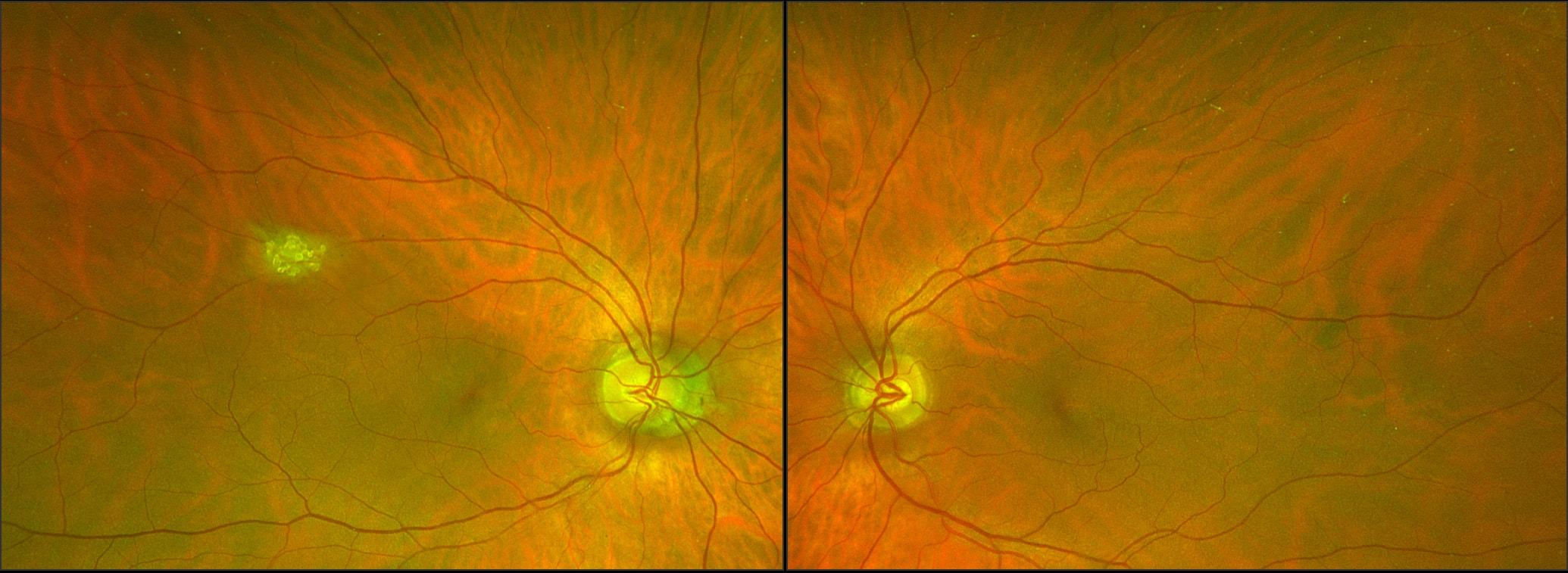
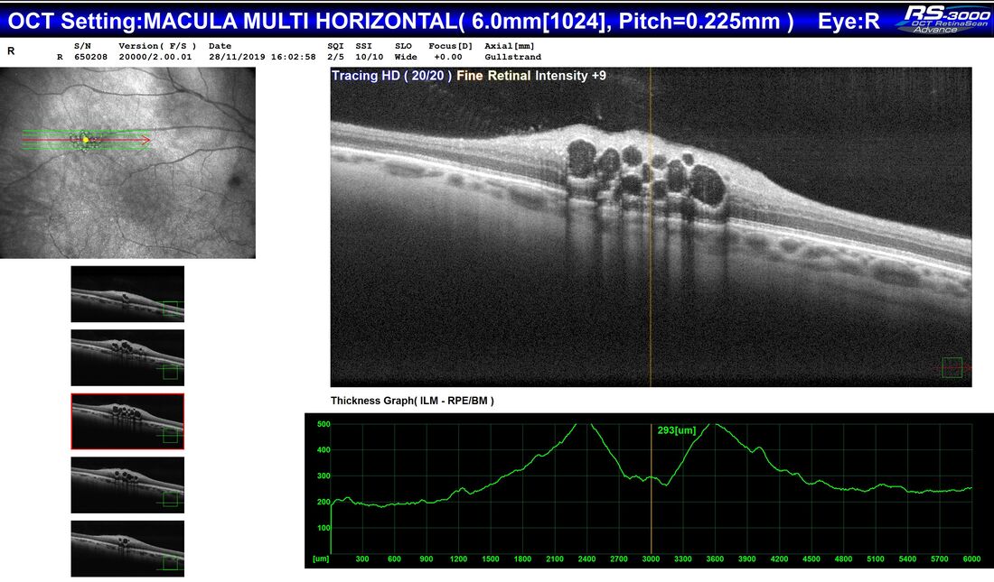
An otherwise well 65 year old man, during a routine examination for spectacles, was found by an Optometrist to have a lesion in the right fundus that had not been previously documented. There was no relevant past history of any ocular or systemic conditions. The lesion appears raised, white and multinodular, with a scalloped edge and patchy calcification - reminiscent of optic nerve head drusen, but in a peripheral location. On OCT, the lesion appears to have a multi cystic structure, each "cyst" consisting of a hyper reflective edge, casting an optical shadow, with an optically empty centre. This is a typical astrocytic hamartoma or "mulberry lesion" - a benign retinal tumour composed of glial cells (connective tissue cells of the nervous system), predominantly astrocytes. It can occur singly or in multiple locations. It is most often associated with tuberous sclerosis (TS) or Bourneville's Disease, but may also be found rarely in patients with neurofibromatosis. Although the finding may point toward a systemic association, it can also be found incidentally on retinal examination as an idiopathic and spontaneous lesion, as in this case.
Apart from recommending the patient have his retina examined periodically in the future, no treatment was required.
0 Comments
A 21 year old male was referred to South West Eye Surgery by his GP with blurred vision and a central visual field defect (a blurred patch) in his right eye. He had awoken 2 days previously with a headache, fatigue and bloodshot eyes.
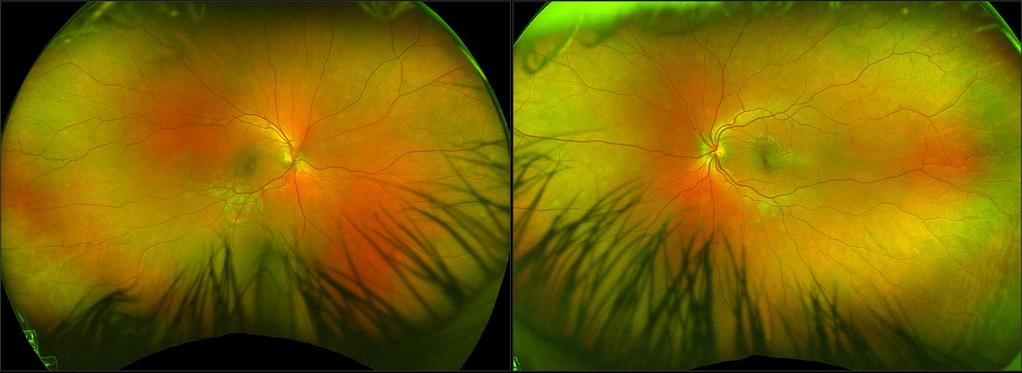
On examination, visual acuities were Right 6/12 and Left 6/5. He had bilateral conjunctival hyperaemia (red eyes), but little anterior or posterior uveitis (internal eye inflammation) of note. |
AuthorDr Vincent Lee, Archives
April 2020
Categories
All
|
Proudly powered by Weebly


 RSS Feed